
EEG representing on brain surface using volume rendering
DOI:
https://doi.org/10.14464/ess.v8i1.476Abstract
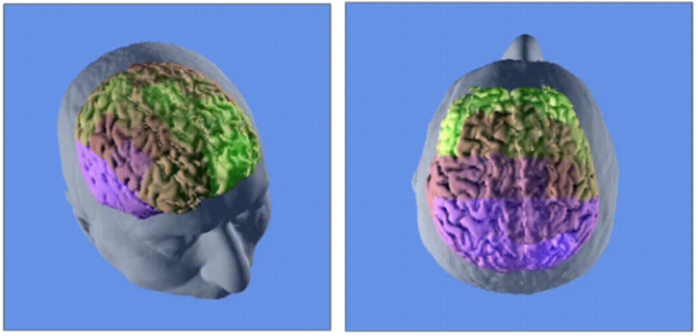
Brain research is challenging. One of the standard research methods is electroencephalography (EEG). As a rule, this study is presented in the form of graphs. This article describes an approach in which this data is mapped onto a brain model generated from a magnetic resonance imaging (MRI) scan. This allows you to look at the EEG study from a different point of view. An MRI scan will also allow you to take into account some of the features of the brain. This is an advantage over mapping just to a brain template. This non-invasive system can be implemented to monitor the patient in real-time, for example, during space flight.
Downloads
Published
Issue
Section
License
Copyright for articles published in this journal is retained by the authors. The content is published under a Creative Commons Licence Attribution 4.0 International (CC BY 4.0). This permits use, distribution, and reproduction in any medium, provided the original work is properly cited, and is otherwise in compliance with the licence.

